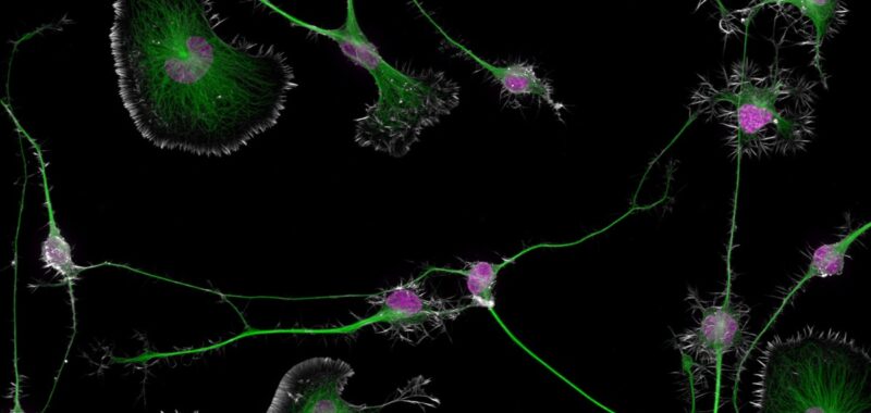
This year’s winning entry arose from Cisterna’s research into a protein crucial for building brain cell structure (profilin 1, or PFN1); that structure is essential for functional cellular transport. He found that when the protein and related processes are disrupted, the microtubule highways can malfunction and cause damage to the cells. Capturing the actin, microtubules, and nuclei with photomicroscopy was a painstaking process that took about three months just to perfect the staining process. Cisterna and Vitriol paid particular attention to getting just the right field of view and got the image they were waiting for after three hours of observation.
“At 50 years, Nikon Small World is more than just an imaging competition—it’s become a gallery that pays tribute to the extraordinary individuals who make it possible,” said Nikon Instruments rep Eric Flem. They are the driving force behind this event, masterfully blending science and art to reveal the wonders of the microscopic world and what we can learn from it to the public. Sometimes, we overlook the tiny details of the world around us. Nikon Small World serves as a reminder to pause, appreciate the power and beauty of the little things, and to cultivate a deeper curiosity to explore and question.”
Here are the remaining top 20 winners of this year’s contest, ranging from close-up views of octopus eggs, green crab spider eyes, and slime molds to capturing the electric arc between a pin and wire, and an insect egg that has been parasitized by a wasp. You can check out the full list of winners, as well as several honorable mentions, here.
And the winners are…
Marcel Clemens/Nikon Small World
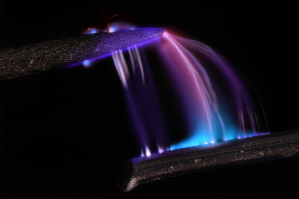
Second place: Electrical arc between a pin and a wire.
Marcel Clemens/Nikon Small World
Chris Romaine/Nikon Small World
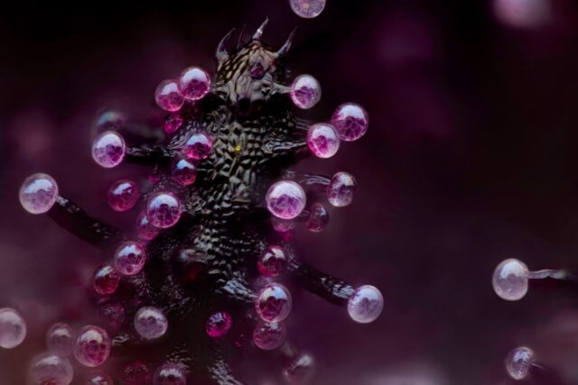
Third place: Leaf of a cannabis plant. The bulbous glands are trichomes. The bubbles inside are cannabinoid vesicles.
Chris Romaine/Nikon Small World
Amy Engevik/Nikon Small World
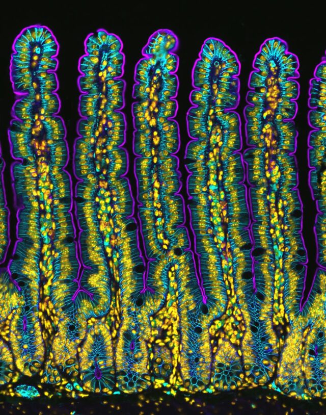
Fourth place: Section of the small intestine of a mouse.
Amy Engevik/Nikon Small World
Third place: Leaf of a cannabis plant. The bulbous glands are trichomes. The bubbles inside are cannabinoid vesicles.
Chris Romaine/Nikon Small World
Fourth place: Section of the small intestine of a mouse.
Amy Engevik/Nikon Small World
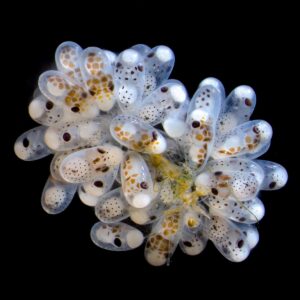
Thomas Barlow & Connor Gibbons/Nikon Small World
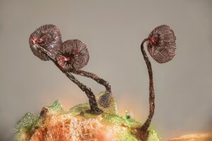
Henri Koskinen/Nikon Small World
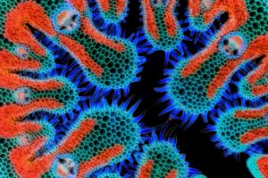
Gerhard Vlcek/Nikon Small World
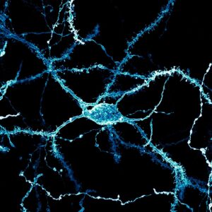
Stephanie Huang/Nikon Small World
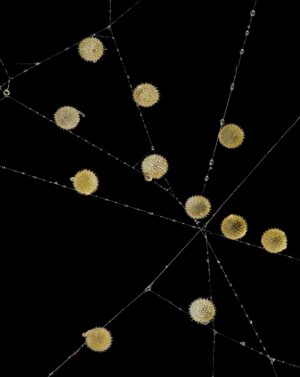
John-Oliver Dunn/Nikon Small World
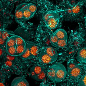
Jan Martinek/Nikon Small World
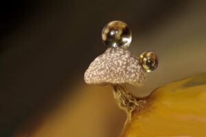
Ferenc Halmos/Nikon Small World
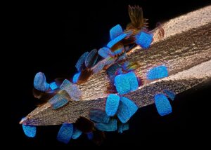
Daniel Knop/Nikon Small World
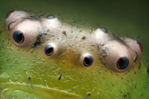
Patel Blachowicz/Nikon Small World
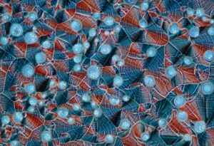
Marek Miś/Nikon Small World
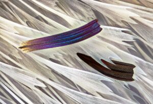
Sébastien Malo/Nikon Small World
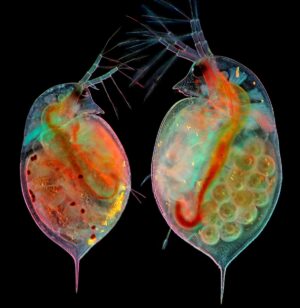
Marek Miś/Nikon Small World
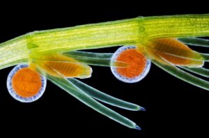
Frantisek Bednar/Nikon Small World
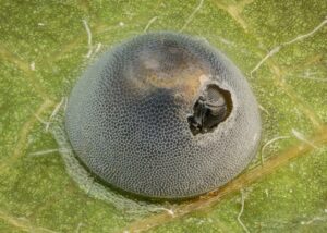
Alison Pollack/Nikon Small World
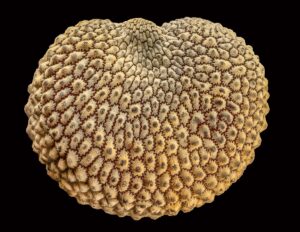
Alison Pollack/Nikon Small World
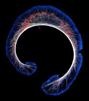
Bruno Cisterna & Eric Vitriol/Nikon Small World

